Why Vitiligo Looks So Different on Dark Skin—And Why Doctors Keep Missing It

Vitiligo is a chronic skin condition that causes the formation of white patches of skin that may appear anywhere on the body. This condition can show up in all races more or less equally, but can be especially more conspicuous—and sometimes more difficult to treat—in darker skin due to the strong color contrast with normal skin. Unfortunately, medical cosmetics and dermatological literature have most often dealt with lighter skin types, leaving both patients and practitioners poorly informed. This article discusses the biology of vitiligo, the way color functions in the skin, and why vitiligo differs in people with darker skin.

What is vitiligo?

Vitiligo is a chronic, autoimmune disease. In other words, the body’s immune system incorrectly attacks the body’s own cells, in this instance called melanocytes. Located in the skin’s outer layer (epidermis), these are cells that have the special function to produce melanin!

Simplified diagram of a melanocyte

Melanin colors the skin, hair, and eyes. Where melanocytes are lost or are not functioning, the areas involved lose their pigment and turn milky white.

Vitiligo is one of two types: non-segmental (more common and occurs on both sides of the body symmetrically) or segmental (less common and is typically on one side of the body). The disease is neither painful nor contagious, but can be devastating to one’s self-esteem.

How Skin Pigmentation Is Formed

In order to find out why vitiligo appears more on darker skin, we have to examine how skin color is created. Melanocytes produce melanin and package it into small organelles known as melanosomes, which are transported to the surrounding skin cells. We all have roughly the same number of melanocytes, but in darker-skinned individuals, the cells are more active and produce more melanin.

When people with darker skin lose pigment, the contrast between the light spots and the rest of the skin is so much more pronounced. In light skin, the spots may be quite light or pink.

Why Vitiligo Appears So Different on Dark Skin

Dark-skin vitiligo patches tend to be very noticeable and snow white, attracting more attention than they would if they were located on light skin. In fact, most dermatology textbooks and instructional photos of skin diseases show vitiligo in light skin. This contributes to the delayed or erroneous diagnosis of the disease in darker patients.

Inequalities in Skin of Color Diagnosis and Treatment

Vitiligo may be misdiagnosed in dark-skinned patients as another skin condition, such as tinea versicolor, post-inflammatory hypopigmentation, or chemical leukoderma. This is particularly so for physicians who are not used to dealing with dark skin. After proper diagnosis, treatment may involve creams with corticosteroids, calcineurin inhibitors (such as tacrolimus), and narrowband UVB phototherapy. Depigmentation therapy may be considered for severe cases. But not everyone chooses to get treated. Most individuals would prefer to accept the way they appear and search for makeup products that will enable them to hide their imperfections. Even then, very few makeup or concealer products are suitable for dark skin.

Closing Comments 

Vitiligo is not riskier on darker skin, but can be readily noticeable, less understood, more frequently, and under-treated or mistreated. Diversity role models, more medical training, and education are the solutions to all of these problems. If you or your relative has vitiligo on darker skin, you need to know that help is growing—and your skin’s narrative needs to be told accurately, sensitively, and with pride.

References

Pietrangelo, Ann. “Vitiligo on Black Skin: What You Should Know.” Medical News Today, Healthline Media, 28 Oct. 2022, https://www.medicalnewstoday.com/articles/vitiligo-black-skin.

Cleveland Clinic. “Vitiligo.” Cleveland Clinic, 13 Oct. 2023, https://my.clevelandclinic.org/health/diseases/12419-vitiligo.

Hitti, Miranda. “What People of Color Should Know about Vitiligo.” WebMD, 6 June 2023, https://www.webmd.com/skin-problems-and-treatments/features/vitiligo-darker-skin-tones.

NHS. “Vitiligo.” NHS, National Health Service UK, 13 Feb. 2024, https://www.nhs.uk/conditions/vitiligo/.

Goldman, Lisa Zamosky. “Vitiligo and People of Color: The Nuances of a Visible Condition.” WebMD, 6 June 2023, https://www.webmd.com/skin-problems-and-treatments/features/vitiligo-poc-nuance.

Washington’s Dermatology Divide Is Leaving Rural Patients Behind

26 Best Small Towns in Washington State with Tons of Character - The  Emerald Palate

If you live in Seattle, getting a weird mole or rash checked out by a dermatologist might just mean a quick trip across town. But if you live in a small town in eastern Washington—or don’t have a car, reliable internet, or time off work—that same appointment could take months to get and hours to reach. For many people in our state, especially in rural areas, skin care just isn’t easy to access. 

And that’s a problem. Skin conditions aren’t always just about appearance—they can be uncomfortable, painful, or even life-threatening. Skin cancer, for example, is one of the most common cancers in the U.S., but it’s also one of the most treatable when caught early. Yet in rural areas, access to dermatologists is alarmingly scarce—with only 0.085 dermatologists per 100,000 people, compared to 4.11 per 100,000 in urban regions across Washington and Oregon. So why are so many Washingtonians still waiting to get care?

Here’s where teledermatology can help. It’s a fancy word, but the idea is simple: it’s a way for skin doctors (dermatologists) to look at rashes, moles, acne, and other skin issues through photos or video calls instead of in-person visits. Patients can send in pictures of their skin problem or meet with a doctor over a video call to get answers without needing to leave their home.

During the COVID-19 pandemic, teledermatology took off out of necessity—and it worked surprisingly well. Many skin issues are visible, which makes them easier to diagnose from a screen than, say, a heart condition. And patients who used telehealth often said they liked it. It saved them gas money, time off work, and long drives just to get care.

But now that we’re out of the emergency phase of the pandemic, we risk losing a good thing. Not everyone in Washington can access teledermatology easily. In some rural towns, there’s no reliable internet. Some insurance plans still don’t cover online dermatology visits the same way they cover in-person ones. And some people—especially older adults—don’t feel confident using video calls or sending pictures from a phone or tablet.

That’s why we need to do more. If Washington wants to make sure everyone has a fair shot at getting skin care, we need to invest in a few key things:

  1. Better internet in rural areas so more people can use telehealth tools.
  2. Fair insurance rules that cover online dermatology visits just like in-person ones.
  3. Community telehealth hubs in places like libraries or clinics, where people can go for private, internet-connected appointments—even if they don’t have the technology at home.
  4. Help for patients who aren’t used to using apps or video calls, so they feel comfortable getting the care they need.

This isn’t just about convenience—it’s about fairness. Everyone deserves to get medical help, whether they live in downtown Seattle or a small farming town. Skin health affects our comfort, our confidence, and sometimes even our lives. No one should have to wait months, drive hours, or skip care because the system makes it too hard.

Teledermatology won’t replace every in-person visit. Sometimes a mole needs to be biopsied, or a condition needs to be seen under special lighting. But for many common issues, remote care can be just as good—and a lot more accessible.

Washington has the chance to lead the way in making teledermatology part of our normal healthcare system. The technology is here. The doctors are willing. The only question is whether we’ll make the changes needed to connect more people to care.

Where you live shouldn’t determine whether you get timely, potentially life-saving care. Washington has the tools to close this gap—starting with teledermatology. Now we just need the will to use them.

Sources:

Serra-García, Marta, et al. Urban versus Rural Utilization of Teledermoscopy in Self‑Skin Examinations: A Cohort Study in Oregon and Washington. Dermatology Reports, vol. 15, 2023. https://www.ncbi.nlm.nih.gov/pmc/articles/PMC10327663/

Rise of Teledermatology in the COVID‑19 Era: A Pan‑World Perspective. Digital Health, 2022. https://pubmed.ncbi.nlm.nih.gov/35154805/

Centers for Disease Control and Prevention. Melanoma of the Skin Statistics. Updated June 10, 2025. https://www.cdc.gov/skin-cancer/statistics/index.html

Botox Explained: How a Neurotoxin Became a Beauty Staple

In 2022, over 9 million Botox injections were given worldwide, making it one of the most popular cosmetic treatments today. Known for its ability to reduce wrinkles and give skin a smoother, younger appearance, Botox is particularly popular among celebrities looking to maintain a youthful appearance. But what exactly is Botox, and how does it work?

What is Botox?

Botulinum toxin, or Botox, is a neurotoxic protein derived from a bacteria known as bacterium Clostridium botulinum. Neurotoxic proteins are substances that disrupt the function of the nervous system. Botulinum toxin works by blocking the release of acetylcholine, a neurotransmitter that plays a critical role in muscle contraction, by interfering with the vesicles responsible for releasing acetylcholine. Located in neurons, vesicles are small sacs that store neurotransmitters, like acetylcholine. Botox targets and breaks down the proteins that help these vesicles release acetylcholine, preventing it from being released into the space between the nerve and the muscle.

This prevention of muscle contraction ultimately leads to reduced wrinkles, fine lines, improved skin texture, and overall a more youthful appearance.

While Botox is best known for its cosmetic benefits, it also provides several other medical benefits. This versatile procedure can also mitigate neck spasms and chronic migraines under the same mechanism. Additionally, Botox is effective in treating excessive sweating (hyperhidrosis). Not only is acetylcholine responsible for muscle contraction, but it also plays a key role in triggering sweat production in sweat glands. Botox can be injected in the affected areas, preventing them from producing excess sweat. Botox injections can also treat lazy eye by relaxing the overactive muscle in the eye—specifically, the extraocular muscle, which controls eye movement. 

Different muscles vary in size and strength, so they require varying amounts of Botox to effectively relax the target muscle. Botox injections are administered in units, with 1 unit being a standardized measure of biological potency.

Where is Botox injected?

Botox can be used wherever facial muscles are creating lines. Here are the most common injection sites:

Forehead – to relax those horizontal lines that show up when you raise your eyebrows. 10-30 units of Botox.

Between eyebrows (glabella) – to soften the vertical lines caused by frowning. 5-15 units of Botox on each side.

Outer corners of eyes (crow’s feet) – to soften the orbicularis oculi muscles at the outer corners of the eyes. 10-15 units of Botox per side.

Around the mouth – to reduce smile lines (nasalabial folds) by pulling the corners of the mouth down. 3-6 units of Botox on each side.

How long does Botox last?

Botox injections generally last 3 to 4 months, depending on the dosage and site of injection. In areas with more muscle activity, like around the eyes or mouth, the effects of Botox will wear off faster compared to areas with less movement, like the forehead.

Side effects of Botox

As we’ve learned, Botox prevents the release of acetylcholine and ultimately muscle contraction. After injection, the targeted muscle remains entirely inactive for the treatment’s duration—typically 3 to 4 months. This prolonged inactivity can cause muscle atrophy, meaning that the muscle will reduce in mass, size, and strength. Some studies have shown that if a patient continues to undergo Botox injections in the long term, their skin might experience increased susceptibility to sun damage, premature aging, and dehydration. Granted, the current research on the long-term effects of Botox remains inconclusive.

Takeaway:

Known for its effective ability to smooth wrinkles by temporarily paralyzing facial muscles, Botox continues to reign as one of the most popular cosmetic treatments. Beyond aesthetics, Botox also treats medical conditions like migraines, excessive sweating, and muscle spasms. However, while generally safe, long-term use may lead to muscle atrophy and potential skin changes. As with any procedure, it’s important to weigh the benefits against possible risks and consult a healthcare provider for personalized care.

Bibliography

“Botox.” MedlinePlus, U.S. National Library of Medicine, 2 Aug. 2023, https://medlineplus.gov/botox.html.

“Vesicle.” NCI Dictionary of Cancer Terms, National Cancer Institute, https://www.cancer.gov/publications/dictionaries/cancer-terms/def/vesicle.

“What Areas Can Be Treated with Botox?” Westlake Dermatology & Cosmetic Surgery, https://www.westlakedermatology.com/blog/what-areas-can-be-treated-with-botox/.

“The Long-Term Effects of Botox: What Research Says.” Your Laser Skin Care, https://www.yourlaserskincare.com/blog/the-long-term-effects-of-botox-what-research-says.

Why Skin of Color Still Gets Misdiagnosed — and How We Can Fix It

13,400+ Black Man At Doctors Office Stock Photos, Pictures & Royalty-Free  Images - iStock

In 2012, Lauryn Taylor—a Black woman—was misdiagnosed with eczema. Another doctor thought it might be vitiligo. Then came guesses like pityriasis alba or macular hypomelanosis. But none of them were right. In reality, Lauryn had skin cancer—and it took nearly 10 years to get the correct diagnosis.

I'm a Black Woman and My Skin Cancer Was Misdiagnosed for Years
Lauryn Taylor


Let that sink in — for nearly a decade, Lauryn lived with a misdiagnosis while her skin cancer went untreated, simply because her condition wasn’t recognized on darker skin.

Misdiagnosis isn’t just a medical error—it’s a public health crisis with endangering consequences. Diagnosis of the wrong condition can delay the proper treatment, allowing the condition to worsen over time. Dermatologists may prescribe particular creams or medications that can cause irreversible damage.

It’s also emotionally draining. Patients may feel frustrated or anxious when their ‘treatments’ are not working, leaving them feeling hopeless and dismayed. In desperation, patients will keep paying and returning for medical consultations and buying the wrong products/treatments. This becomes a heavy financial burden.

These are licensed physicians, right? Why are so many of them misdiagnosing patients?

“Probably no doctor is intending to do worse on any type of person, but it might be the fact that you don’t have all the knowledge and the experience, and therefore on certain groups of people, you might do worse,” says Northwestern University professor Matt Groh. In fact, it’s not the dermatologists’ fault — it’s the education system. 

“A large part of dermatology education involves visual recognition,” states dermatologist Jasmine Onyeka Obioha, MD. “Physicians must identify conditions from photos of skin in textbooks or shared in the classroom. But most of the patients in those images are white and only 4.5% of the images show dark skin.” As a result, most medical students don’t know how to identify the skin conditions of patients of color, leading to a disparity in misdiagnosis among racially and ethnically diverse populations. 

However, the misrepresentation of racially/ethnically diverse populations extends beyond dermatology textbooks, reflecting a broader, systemic issue within clinical education and diagnostic training. “Women and racial and ethnic minorities are 20% to 30% more likely than white men to experience a misdiagnosis,” said Johns Hopkins professor of neurology David Newman-Toker. This astounding statistic reveals the racial/ethnic disparities embedded in the foundations of medical training. 

It’s no wonder why physicians often struggle to diagnose conditions in patients whose appearances and symptoms fall outside of the narrow standards they were taught.

What can we do?
In order for us to promote racial equality in dermatology/health care and prevent misdiagnoses, we must:


Reform medical education:
As only ~4.5% of dermatology images feature darker skin, we need to bring that number up to at least 30%. This number ensures equitable inclusion and representation of racially and ethnically diverse populations. 

We need to incorporate modules tailored to diagnosing and treating conditions presented by racially and ethnically diverse populations into the medical curriculum.

Advocate for change:
Acknowledging the issue and the devastating statistics is the first step. Now, to turn awareness into action, we must collectively raise our voices, educate others, and advocate for meaningful change. People of color deserve to be treated with dignity and equity in the healthcare system.

Available resources:
https://guides.ucsf.edu/c.php?g=1081119&p=9159811

https://skinofcolorsociety.org/
https://www.aad.org/member/career/diversity

Bibliography:


1. UCSF Library. Health Disparities and Skin of Color in Dermatology. University of California San Francisco, 2022, https://guides.ucsf.edu/c.php?g=1081119&p=9159811.

2 Skin of Color Society. Home. https://skinofcolorsociety.org/.

3. American Academy of Dermatology. Diversity, Equity, and Inclusion in Dermatology. https://www.aad.org/member/career/diversity.

4.  Miller, Korin. “Doctors Told Me My Skin Cancer Was Eczema for Almost a Decade.” Prevention, 28 May 2021, https://www.prevention.com/health/health-conditions/a36230501/mycosis-fungoides-misdiagnosis/.

5. Kim, Sophia, et al. “Diagnostic Performance of Dermatologists and General Practitioners in Skin Diseases on Diverse Skin Tones.” Nature Medicine, vol. 29, 2023, pp. 1941–1948. https://www.nature.com/articles/s41591-023-02728-3.

6. Loftus, Peter. “Misdiagnosed: Women and Minorities Face Higher Risk of Harm from Medical Errors.” KFF Health News, 15 Dec. 2023, https://kffhealthnews.org/news/article/medical-misdiagnosis-women-minorities-health-care-bias/.

7. Cedars-Sinai. “Why Skin Issues in Patients of Color Are Neglected, Mistreated.” Cedars-Sinai Newsroom, https://www.cedars-sinai.org/newsroom/why-skin-issues-in-patients-of-color-are-neglected-mistreated/.

8. Pew Research Center. Facts About the U.S. Black Population. 9 Feb. 2023, https://www.pewresearch.org/race-and-ethnicity/fact-sheet/facts-about-the-us-black-population/.

9. Massachusetts General Hospital. Skin Assessment in Patients with Dark Skin Tone. Munn Center, Apr. 2023, https://www.mghpcs.org/MunnCenter/Documents/weekly/apr-23/Skin-Assessment-in-Patients-with-Dark-Skin-Tone.pdf.

10. Trafton, Anne. “Doctors Struggle to Diagnose Diseases in Patients with Darker Skin.” MIT News, 5 Feb. 2024, https://news.mit.edu/2024/doctors-more-difficulty-diagnosing-diseases-images-darker-skin-0205.

11. TEDx Talks. “Why Doctors Misdiagnose Skin Conditions on Darker Skin.” YouTube, uploaded by TEDx, 7 Oct. 2022, https://www.youtube.com/watch?v=2JUQo-PnY2g.

Biochemical Markers of Psoriasis: What’s Happening Beneath the Skin?

This image has an empty alt attribute; its file name is AD_4nXd2JzncBsi_DIKtCZDMhchlrVumhV5XgKdTVUUtag3hoxQrdZ5x-ItRsFAWX3tXcI9E9fX4GTp-ELrNHPcumBTB1v_HmIBRpB6aIuSIetURY7nHsdR7kiWXR991T8mID5DjBWEZ

Often confused with atopic dermatitis, psoriasis is also an incurable chronic skin condition that is characterized by itchy/inflamed and scaly patches of skin. As psoriasis affects ~2% of the United States population, the demand for treatment continues to soar. Unfortunately, psoriasis is a complex autoimmune condition that entails various factors. In this post, we’ll learn about the causes of psoriasis, the role of the immune system in psoriasis, remedies, as well as how it differs from other inflammatory skin conditions like eczema.

What causes psoriasis?

The root causes of psoriasis start with genetics and environmental triggers. This combination is integral for psoriasis, with genetic predispositions endangering the host to an overreactive immune response, which can be provoked by an environmental trigger. Triggers include skin injuries, infections, stress, alcohol, and more.

What happens during a psoriasis flare-up?

Once triggered, the immune system begins to malfunction. Crucial immune cells known as t cells—responsible for fighting off pathogens—become overactive. They begin to mistakenly orchestrate an attack on skin cells, assuming them to be pathogens. First, T cells release cytokines (typically IL-17 and IL-23)—proteins that regulate immune responses and contribute to inflammation. These cytokines then stimulate the overproduction of keratinocytes (skin cells) to try to repair the damage. The basal layer of the epidermis (where keratinocytes are produced) then goes overdrive, resulting in rapid cell division. This manifests as flaky, inflamed, and itchy lesions of the skin of psoriasis. 

Here is a flowchart to help demonstrate the process:


Genetic predisposition and environmental triggers 

T cells become overactive

T cells release cytokines

 Cytokines stimulate keratinocyte overproduction

Visible symptoms of psoriasis (Scaly, itchy plaques)

Psoriasis vs. atopic dermatitis

Psoriasis and atopic dermatitis are both chronic skin conditions that manifest as flaky, inflamed, and itchy patches of skin. However, the difference between these two comes down to their underlying cause. As we’ve learned, psoriasis is an autoimmune condition that involves the immune system mistakenly attacking the body. In contrast, atopic dermatitis is an inflammatory condition involving a defective/hypersensitive skin barrier due to genetic mutation. Although psoriasis can also be triggered by environmental triggers, think of atopic dermatitis as being the direct, inflammatory response to a trigger. Psoriasis, on the other hand, happens because the immune system is already defective and attacking the body’s own skin cells, and outside triggers just make it worse.

Types of psoriasis

Psoriasis can take on several forms, depending on the types of cytokines involved. Accounting for 80-90% of psoriasis cases, plaque psoriasis is the most common form. Primary cytokines involved: IL-17 and IL-23. Manifests as raised, silver-colored, scaly patches of skin. Most commonly forms on the elbows and knees. Psoriasis can also take several other forms, affecting different parts of the body. These include: guttate psoriasis (torso, upper arms, and legs), pustular psoriasis (palms of hands or soles of feet), inverse psoriasis (skin folds), and many more.

Plaque psoriasis

Treatments:

Luckily, scientists have developed various treatment options for psoriasis that work by reducing inflammation and slowing the overproduction of skin cells. Treatments include topical therapies (creams and ointments), light therapy, and oral or injected medications. The best option for each patient depends on the severity of the condition, medical history, and lifestyle.

Conclusion:

Understanding the biochemistry and immunology behind psoriasis reveals the complexity of this abstract, chronic, autoimmune condition. Although there isn’t currently a direct cure for psoriasis, several treatments have been developed to mitigate symptoms, reduce inflammation, and prevent flare-ups. The more we understand the underlying cause of psoriasis, the closer we get to hopefully curing this chronic condition.

Bibliography

American Academy of Dermatology Association. “What Causes Psoriasis?” AAD, https://www.aad.org/public/diseases/psoriasis/what/causes. Accessed 13 Apr. 2025.

Rendon, Adriana, and Benjamin Schäkel. “Psoriasis Pathogenesis and Treatment.” International Journal of Molecular Sciences, vol. 19, no. 5, 2019, p. 1475, https://pmc.ncbi.nlm.nih.gov/articles/PMC5751129/. Accessed 13 Apr. 2025.

National Institute of Arthritis and Musculoskeletal and Skin Diseases. “Psoriasis.” NIAMS, U.S. Department of Health and Human Services, https://www.niams.nih.gov/health-topics/psoriasis. Accessed 13 Apr. 2025.

“Psoriasis.” TeachMe Paediatrics, https://teachmepaediatrics.com/dermatology/dermatology/psoriasis/. Accessed 13 Apr. 2025.

Greb, Jennifer E., et al. “Psoriasis.” International Journal of Molecular Sciences, vol. 20, no. 10, 2019, p. 2558, https://www.mdpi.com/1422-0067/20/10/2558. Accessed 13 Apr. 2025.

Radiant Dermatology. “Psoriasis.” Radiant Dermatology & Skin Cancer Center, https://www.radiantdermtx.com/view-medical-dermatology/psoriasis. Accessed 13 Apr. 2025.

Thomas, Liji. “What Are T Cells?” News Medical, 27 Feb. 2019, https://www.news-medical.net/health/What-are-T-Cells.aspx. Accessed 13 Apr. 2025.

Di Meglio, Paola, et al. “The Role of APCs in Psoriasis Pathogenesis.” Frontiers in Immunology, vol. 6, 2015, https://pmc.ncbi.nlm.nih.gov/articles/PMC4437803/. Accessed 13 Apr. 2025.

WebMD. “Cytokines and Psoriasis.” WebMD, https://www.webmd.com/skin-problems-and-treatments/psoriasis/cytokines-psoriasis. Accessed 13 Apr. 2025.

Parisi, Rosalind, et al. “Psoriasis Vulgaris: A Comprehensive Overview.” Dermatology and Therapy, vol. 10, no. 2, 2020, pp. 261–273, https://pmc.ncbi.nlm.nih.gov/articles/PMC7122924/. Accessed 13 Apr. 2025.

Tyrosinase Inhibitors vs. Melanogenesis Inhibitors: Which is Better for Treating Pigmentation?

What is pigmentation? - CellDerma

What causes pigmentation?

Responsible for providing color to our skin, hair, and eyes, melanin is an essential, naturally produced pigment that our body–and most organisms— is constantly producing. Our natural skin tone is largely accredited to our genetically determined melanin production rate, which vastly varies across ethnicities. Inhabitants of regions with more intense UV radiation have evolved to develop genetic mutations that increase their melanin production and thus have darker skin tones, whereas people in lower UV regions have lighter skin tones. The process of melanin production—otherwise known as melanogenesis—entails the conversion of tyrosinase—a natural enzyme—into melanin. 

Simplified pathways of tyrosine-derived melanin synthesis showing... |  Download Scientific Diagram
Process of Melanin Synthesis
(ResearchGate)

How does UV radiation influence melanin production?

As a defense mechanism against UV radiation, the skin initiates melanogenesis. Melanin then transfers to keratinocytes—the cell that produces keratin, an essential protein in the epidermis—where the melanin internally absorbs UV rays and reduces DNA damage. However, if the skin receives excessive exposure to UV radiation—without sun protection—the melanin will eventually fail to provide skin protection, leading to sun damage and, soon enough, hyperpigmentation. Although tyrosinase production is also largely influenced by genetics and exposure to UV radiation, tyrosinase, and melanin production can be reduced by tyrosinase inhibitors.

What are tyrosinase inhibitors?

Whether found synthetically in ingredients like azelaic acid or naturally in ingredients like aloe vera, tyrosinase inhibitors are widely used to even out skin tone by blocking tyrosinase activity during catalytic reaction, thereby reducing melanin production and, in some cases, breaking down and degrading pre-existing melanin. Their versatility makes them a key component in the treatment of hyperpigmentation, melasma, and age spots. While different tyrosinase inhibitors function differently, they ultimately yield the same results—reduced melanin synthesis and a more even complexion. Here are some examples of tyrosinase inhibitors with varying potency.

Niacinamide: Although not technically a tyrosinase inhibitor, niacinamide Inhibits melanosome transfer—the process of turning melanin into a visible pigment. Niacinamide does not alter tyrosinase production, making it a more gentle alternative to typical tyrosinase inhibitors. It can be applied up to twice daily.


Azelaic acid: Inhibits tyrosinase and gently reduces pigmentation by targeting hyperactive melanocytes—cells that produce melanin. Azelaic acid is slightly more potent than niacinamide. It can be applied up to twice daily.

Hydroquinone: Directly inhibits tyrosinase and melanin synthesis. It can be used up to twice a day for 3-6 months as treatment for intense hyperpigmentation or melasma; to avoid irritation, it should not be used any longer, as it is highly potent.

Melanogenesis inhibitors are often confused with tyrosinase inhibitors, and although they yield similar results—reducing pigmentation—tyrosinase inhibitors are a subset of melanogenesis inhibitors, which differ in that they affect melanin synthesis as a whole rather than directly targeting tyrosinase. Alongside just targeting tyrosinase production, melanogenesis inhibitors also promote melanin degradation and target melanogenesis pathways, blocking melanin from transferring to keratinocytes.

Which inhibitor is better?

Although melanogenesis inhibitors target several steps in melanogenesis as opposed to directly targeting tyrosinase activity—as seen with tyrosinase inhibitors—that doesn’t necessarily mean that one is stronger or more effective than the other. In general, melanogenesis inhibitors are preferred for more gradual and long-term treatment for pigmentation, whereas tyrosinase inhibitors are stronger for more rapid pigmentation treatment and results. While both are effective in their own ways, combining these treatments with sun protection, exfoliants, and antioxidants ensures the best results for maintaining an even skin tone and treating hyperpigmentation.

Bibliography

Boissy, R. E. “Melanosome Transfer to and Translocation in the Keratinocyte.” Experimental Dermatology, vol. 7, no. 3, 1998, pp. 143-150. PubMed.

National Cancer Institute. “Melanocyte.” NCI Dictionary of Cancer Terms, n.d., [https://www.cancer.gov/publications/dictionaries/cancer-terms/def/melanocyte](https://www.cancer.gov/publications/dictionaries/cancer-terms/def/melanocyte#:~:text=(meh%2DLAN%2Doh%2D,contains%20the%20pigment%20called%20melanin.).

University of Rochester Medical Center. “What Is Skin Pigmentation?” Health Encyclopedia, n.d., https://www.urmc.rochester.edu/encyclopedia/content?contenttypeid=85&contentid=p01359.

Costin, G. E., and V. J. Hearing. “Human Skin Pigmentation: Melanocytes Modulate Skin Color in Response to Stress.” The FASEB Journal, vol. 21, no. 4, 2007, pp. 976-994. PMC.

Burkhart, C. G., and H. R. Burkhart. “Hydroquinone.” StatPearls, StatPearls Publishing, 2019. NCBI.

Pillaiyar, T., M. Manickam, and V. Namasivayam. “Skin Whitening Agents: Medicinal Chemistry Perspective of Tyrosinase Inhibitors.” Journal of Enzyme Inhibition and Medicinal Chemistry, vol. 32, no. 1, 2017, pp. 403-425. PMC.

Chang, T. S. “Natural Melanogenesis Inhibitors Acting Through the Down-Regulation of Tyrosinase Activity.” International Journal of Molecular Sciences, vol. 10, no. 6, 2009, pp. 2440-2475. PMC.

Sasaki, M., K. Hasegawa, and Y. Takahashi. “Recent Advances in Skin Lightening Agents: A Comprehensive Review.” Journal of Dermatological Science, vol. 110, no. 1, 2023, pp. 12-24. PMC.

What Happens During an Eczema Flare-Up? A Scientific Breakdown


Eczema Symptoms | Clovis Dermatology

What is eczema?
Eczema is a common skin condition characterized by inflamed, itchy patches of dry and scaly skin. This chronic condition affects roughly 10% of the US population. These patches result from a combination of environmental triggers and immune system reactions, often with a genetic predisposition. There are several types of eczema, including atopic dermatitis—the most common type—and contact dermatitis, among others. The key difference between atopic dermatitis and contact dermatitis comes down to the underlying cause and immune response. Atopic dermatitis is a chronic condition that can be triggered by both external factors and internal influences, such as stress, but is ultimately caused by genetic and immune system factors. In contrast, contact dermatitis is an acute condition that is directly caused by irritants like soaps or chemicals and can easily be prevented by avoiding said irritants.

Who suffers from atopic dermatitis?
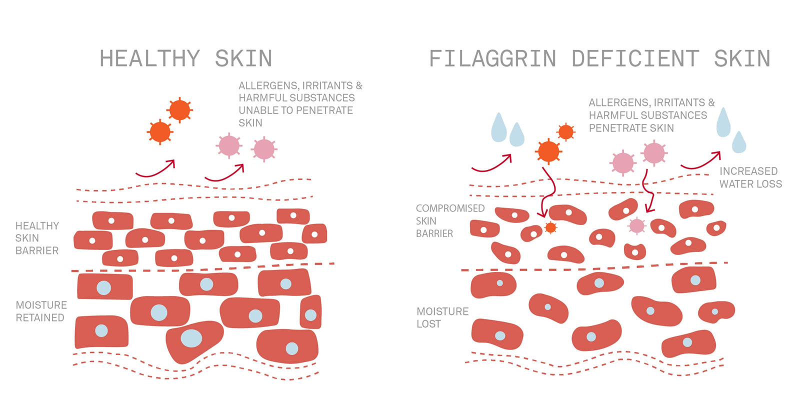
The integration of genetic mutations and a hyperactive immune response in individuals with atopic dermatitis leads to persistent eczema flare-ups. In particular, these individuals have a loss-of-function genetic mutation in their filaggrin gene—one of 70 genes comprising the Epidermal Differentiation Complex—which is crucial for maintaining the epidermis and protecting it from allergens and irritants. This genetic mutation causes the affected individual to produce less filaggrin protein, ultimately resulting in a compromised skin barrier and increased susceptibility to environmental triggers. This single filaggrin defect leads to a cascade of dysfunctions in the body, including reduced natural moisturizing factor levels and a hyperactive immune system.

Filaggrin and eczema – Nursem Skincare
(https://www.nursem.co.uk/pages/filaggrin-and-eczema)

What happens during a flare-up?
An atopic dermatitis flare-up is a multi-step process that begins with a trigger—environmental factors, irritants, allergens, etc. Because the skin barrier of these individuals is compromised and more susceptible to infiltration, allergens and irritants can penetrate more easily. Once the trigger reaches the compromised skin barrier, antigen-presenting cells capture the trigger and carry it over to the local lymph nodes. Lymph nodes are organs that house various immune cells that help coordinate the immune response. T cells – a key type of immune cell – activate and then identify the trigger to initiate the release of cytokines—signaling proteins that—when overproduced—are primarily responsible for the redness and inflammation associated with atopic dermatitis.

How do topical medications alleviate flare-ups?

The complex combination of genetics, environmental triggers, and the immune system makes eczema uncurable, although its symptoms can be mitigated. Topical steroids and inhibitors are commonly used to manage inflammation in conditions like atopic dermatitis. Topical steroids are applied directly to the skin, where they enter cells and bind to receptors in the cytoplasm. This binding triggers the formation of a steroid-receptor complex, which then migrates to the nucleus. In the nucleus, the complex binds to and modifies specific DNA sequences, including the one to reduce the production of inflammatory cytokines. This process helps alleviate redness, swelling, and other symptoms of inflammation.
On the other hand, topical inhibitors reduce inflammation by binding to a protein in T cells, creating a complex that inhibits calcineurin, an enzyme responsible for promoting cytokine transcription and T cell activation. Although these topical treatments yield similar results, their function is very different, so prescription of either should be done by a healthcare professional.

Bibliography

National Center for Biotechnology Information. “Chemical Bonding and Molecular Structure.” NCBI Bookshelf, 2019, https://www.ncbi.nlm.nih.gov/books/NBK538209/.

Wang, Y., et al. “Molecular Insights into Chemical Interactions.” PLOS Computational Biology, vol. 17, no. 2, 2021, e1008623. https://pmc.ncbi.nlm.nih.gov/articles/PMC7880084/.

“—.” American Cancer Society, www.cancer.org/cancer/diagnosis-staging/lymph-nodes-and-cancer.html#:~:text=Lymph%20vessels%20send%20lymph%20fluid,filter%20fluid%20in%20those%20areas.

National Eczema Association. “Eczema Topical Treatments | National Eczema Association.” National Eczema Association, 4 Mar. 2022, nationaleczema.org/eczema/treatment/topicals.

Debunked: Are Beef Tallow Moisturizers Better than Traditional Moisturizers?

Applying beef tallow – rendered fat of a cow – on the skin has an extensive historical background, dating back centuries ago, and has recently regained popularity on social media, particularly in the form of a moisturizer. Unlike traditional moisturizers that use glycerin or hyaluronic acid to moisturize the skin, beef tallow-based moisturizers use solely beef tallow and oftentimes, oil. I decided to look into beef tallow-based moisturizers to determine whether or not they are effective/safe, and how they compare to traditional moisturizers.


The skin benefits of beef tallow

Beef tallow moisturizers are promoted to deeply moisturize the skin and soothe acne, however, there’s insufficient research to support this claim. As stated by the NCBI, more research is still needed for beef tallow to be used as a cosmetic product for humans. The potential benefits of beef tallow for the skin are as follows: beef tallow’s rich content of fatty acids is assumed to protect the skin barrier from pollutants/inflammation, thus promoting a stronger lipid barrier. Fatty acids also help retain water/moisture in the skin, preventing a process known as transepidermal water loss, which occurs when water evaporates from the skin, drying it out. Beef tallow advocates claim that the fatty acids replicate the skin’s natural sebum, making it a “better”  alternative than traditional moisturizers.

The drawbacks of beef tallow

Despite the moisturizing benefits of fatty acids, the chemical construction of fatty acids makes them comedogenic and not fungal acne-safe; the rich content of fatty acids and the thick nature of beef tallow may feel thick and heavy on the skin. Moreover, beef tallow moisturizers typically contain oil – often olive oil – alongside beef tallow. This makes beef tallow moisturizers comedogenic, alongside feeling heavy and greasy, with a rating of 2 out of 5 on the comedogenic scale. For this reason, beef tallow moisturizers are not advised for individuals with acne-prone skin despite companies claiming that they’re suitable for all skin types. However, for individuals with dry skin or eczema, beef tallow moisturizers may be tolerable.

Comedogenic Scale


What do reviews say about beef tallow moisturizers?

Due to the lack of research regarding beef tallow for the skin, I looked into reviews from customers. One customer experimented with a beef tallow moisturizer for one week and found that the product was heavy and left a greasy finish. By day five, her face started breaking out – this likely alludes to the comedogenic formulations of beef tallow moisturizers. While this customer had a poor experience with beef tallow, it should be noted that her skin type may not be compatible with these thick moisturizers, as she doesn’t have super dry or eczema-prone skin. 

For some reason, social media has tried to label traditional moisturizers as having “dangerous chemicals,” scaring the public into buying these natural, beef tallow-based moisturizers. Although some customers may have a pleasant experience with these products, there’s still insufficient scientific evidence/research to back up using beef tallow for the skin, so it would be safe to use traditional moisturizers instead. The ingredients and nature of beef tallow moisturizers make them likely heavy and comedogenic for acne-prone individuals, as proven by customer reviews. As long as you invest in a quality product that doesn’t have parabens, sulfates, etc., traditional moisturizers are completely safe and effective.

Works Cited

“Does Tallow Clog Pores? The Comedogenic Scale Explained.” Sun & Moo, sunandmoo.com/blogs/sun-moo/does-tallow-clog-pores-the-comedogenic-scale-explained. Accessed 16 Jan. 2025.

Holender, Samantha. “So, People Are Slathering Their Faces In Beef Tallow Now?” Vogue, www.vogue.com/article/beef-tallow-for-skin. Accessed 16 Jan. 2025.

Medaris, Anna. “Beef Tallow for Skin: What Dermatologists Think About the Trend.” Edited by Ross Radusky. Everyday Health, 29 Feb. 2024, www.everydayhealth.com/healthy-skin/beef-tallow-for-skin-what-dermatologists-think-about-the-trend/#:~:text=The%20Takeaway,irritation%20rather%20than%20any%20benefits. Accessed 16 Jan. 2025.

“Relative irritancy of free fatty acids of different chain length.” National Library of Medicine, pubmed.ncbi.nlm.nih.gov/233889/. Accessed 16 Jan. 2025.”Tallow, Rendered Animal Fat, and Its Biocompatibility With Skin: A Scoping Review.” National Library of Medicine, pubmed.ncbi.nlm.nih.gov/38910727/. Accessed 16 Jan. 2025.