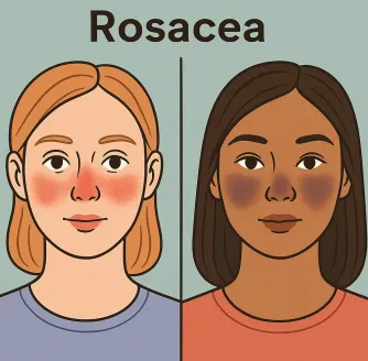
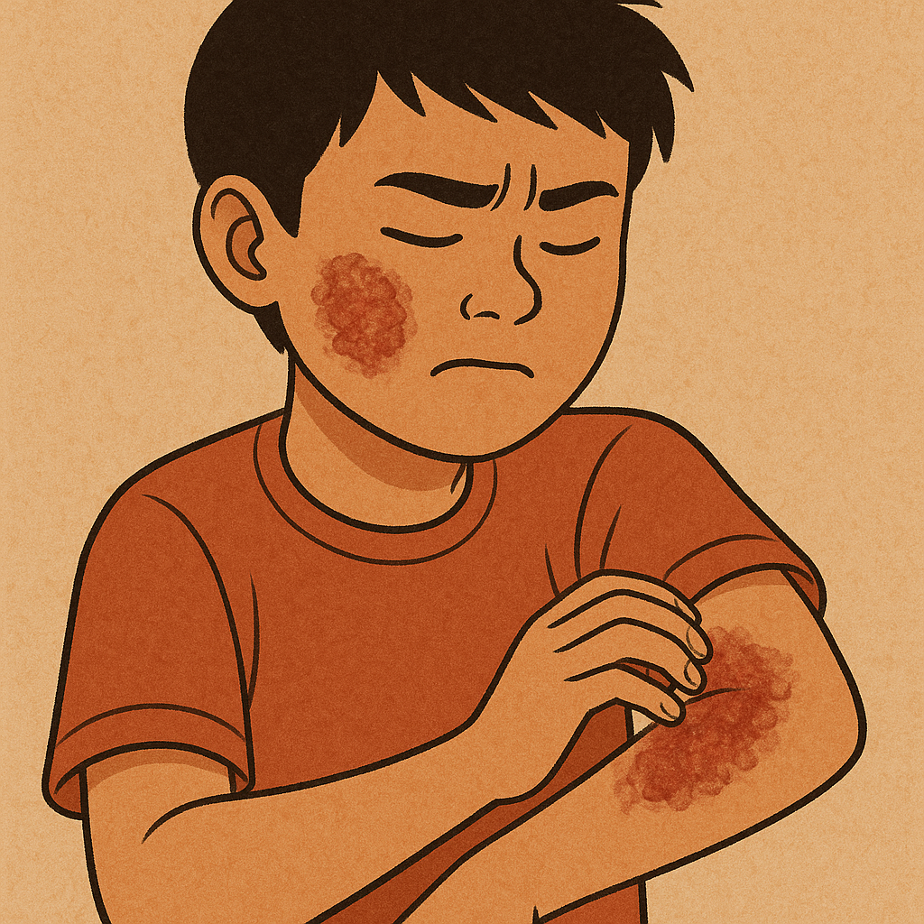
Stratum Corneum Thickness Explained: Why It Matters for Skin of Color

Our skin is incredibly adaptive. Depending on your ethnicity and the environment you live in, the way your skin looks and functions can vary quite a bit from someone else’s. One part of the skin where this really shows is in the stratum corneum.

First, what exactly is the stratum corneum?

You might already know that skin has three main layers: the hypodermis, the dermis, and the epidermis. The epidermis is the outermost layer, and it’s made up of several sublayers. Sitting right at the top is the stratum corneum.

Even though it is the thinnest layer, it still has the main function of the skin to keep kept healthy. It works as a protective barrier, helps with water retention, and protects the skin against various types of environmental stress. What is interesting is how the stratum corneum can be very different in terms of thickness and structure between people and even more so among various ethnic groups. These differences are not just skin-deep—they can impact things like how well your skin holds hydration, to the extent it may become reactive to inflammation or skincare products.

Why thickness matters

The stratum corneum is not identical in everyone. Studies indicate that the skin of East Asians normally has a stratum corneum that is not only thicker but also more compact in comparison with the strata of people with lighter skin. While this sounds like a positive aspect, and it is to some extent (a stronger barrier may prevent irritants from entering), it also implies that the skin may lose its water very fast. This can make the skin dry, and the occurrence of eczema can increase. Besides, Asian skin is prone to post-inflammatory hyperpigmentation (PIH), after the event of a minor breakout or irritation, a dark spot is the aftermath that stays for a long time.

In the same manner, African skin usually has a stratum corneum that is thicker and more compact. This gives the skin extra protection from external traumas, but the mentioned benefits come with some of the same problems. The skin is capable of losing water very fast which is one of the causes of dryness and also the condition which many people know as “ashy”. Hyperpigmentation is yet another issue that occurs frequently, and the reason is that even a slight irritation may result in dark marks that are indistinguishable from the skin and are there long after the skin has recovered.

On the contrary, Caucasian skin, in general, has a relatively thin stratum corneum. In this case, skincare products can penetrate the skin quite easily, and the effects can be seen quicker but at the same time, the skin is more susceptible to environmental stress. The thinner the barrier, the less protective it is, which is the main reason why lighter skin is more highly predisposed to sunburn and certain types of skin cancer.

Why it all matters for your skincare routine

Even if the stratum corneum is measured in microns, it is still the one that controls your skin's behavior and the way your skin reacts to the products. The product penetration is slowed down by a thick barrier, while a thinner one may indicate that the skin is more sensitive. This is the reason why knowing the structure of your skin is very important in the correct ways of treating it.

Takeaway

Of course, no matter what skin type and shade you have, healthy skin starts with taking good care of your skin barrier. This means regular use of emollients, gentle exfoliation, and avoiding the use of harsh products that may be doing more damage than good. The stratum corneum is different from person to person, and understanding this is explains why there is no such thing as one-size-fits-all skincare.

References

Stratum Corneum. ScienceDirect, Topics in Agricultural and Biological Sciences, Elsevier, https://www.sciencedirect.com/topics/agricultural-and-biological-sciences/stratum-corneum.


Murphrey, Morgan B., et al. “Histology, Stratum Corneum.” StatPearls, edited by StatPearls Publishing, https://www.ncbi.nlm.nih.gov/books/NBK513299/.


Rawlings, A. V. “Ethnic Skin Types: Are There Differences in Skin Structure and Function?” International Journal of Cosmetic Science, vol. 28, no. 2, pp. 79–93. PubMed, https://pubmed.ncbi.nlm.nih.gov/18492142/.

Your Water Could Be Ruining Your Skin

When we think about skincare, most of us go straight to products. We obsess over finding the right serum, the perfect cleanser, or moisturizer everyone on TikTok is raving about (like beef tallow…). But here’s something we often overlook: the water coming out of your faucet. Yes, your water. The truth is, your water might be quietly sabotaging your skin, and the effects can be dangerous, varying depending on where you live.

Well, why is this?

Hard water. It’s basically water that has a high mineral content, usually calcium and magnesium. On your skin, they can do more harm than good. If you combine hard water with your soap or cleanser, it doesn’t rinse off as well. Instead, it leaves behind this soapy/slimy residue that can clog pores and cause your skin to feel tight or dry. In the long term, that gunk interferes with your skin barrier. If you have conditions like eczema or acne, that interference can totally make flare-ups even worse. Additionally, leftover minerals can even interfere with the active ingredients in your products, making them less effective. Think of it like trying to spread lotion over sandpaper—your skin just can’t absorb things the same way.

 One quick way I check for hard water is by noticing how products lather. As you rub your cleanser, or even hand soap, and you notice it feels slimy in your hands and doesn’t foam as well as it usually does, that’s a sign the water might be hard. And if you ever travel and suddenly notice your skin acting up, it’s worth checking the local water hardness through a quick Google search. Here’s a handy chart illustrating water pressures across states in the US.

Now here’s where the conversation gets bigger than just skincare tips. Water quality and access are not the same everywhere. Hard water is more common in certain geographic regions, but it’s also more likely to affect communities with aging or underfunded infrastructure. In some poor neighborhoods, individuals just can’t afford to equip their homes with water softeners or quality shower filters that wealthier communities take for granted. Even in the same city, plumbing and maintenance variations may lead to some households receiving consistently harsher water than others.

For individuals of color, these issues are even bigger. Eczema, hyperpigmentation, or scalp problems might not appear the same on darker skin, so they’re dismissed or treated improperly. Add hard water to the mix—exacerbating dryness and redness—and it’s even worse. But still, you never really hear water quality discussed in skincare advice or even in dermatology recommendations. This silence shows that the larger environmental and systemic issues impacting marginalized groups tend to get swept under the rug in health discussions.

So what can you do if you feel your water is part of the solution? The good news is that several low-cost measures can help. Shower filters will reduce mineral content and although some models are expensive, there are cheaper ones that work. For people who have very sensitive skin, applying a rinse with micellar water or even a splash of distilled water makes the residue more loosenable. Selecting gentle, pH-balanced cleansers (and not foamy ones) also makes your skin less susceptible to the harsh effects of hard water. And something easy everyone can do: apply moisturizer as soon as possible after showering to trap in the moisture before minerals and evaporation dry out your skin. The best choice in the long term would ultimately be to install a water softener system in your whole house, but those are quite expensive, costing thousands. 

The bottom line? Skincare isn’t just about your products. Fresh, skin-friendly water should be the standard for all people, no matter where they live. But now it’s a luxury. Our products do matter a lot—yes. But so do the subtle environmental factors that influence our skin health every night. By learning how something as prevalent as water pressure or mineral levels impacts us, not only do we better care for our own skin, but we come to realize why having access to healthy, safe water is a matter of equity, not a luxury. 

How is your water?

References

H₂O Distributors. Hard-Water Map. H₂O Distributors, n.d., https://www.h2odistributors.com/info/hard-water-map/. Accessed 24 Aug. 2025.

U.S. Geological Survey. Hardness of Water. Water Science School, U.S. Geological Survey, n.d., https://www.usgs.gov/water-science-school/science/hardness-water. Accessed 24 Aug. 2025.

Roland, James. “Hard Water vs. Soft Water: Which One Is Healthier?” Healthline, reviewed by J. Keith Fisher, MD, 30 July 2019, https://www.healthline.com/health/hard-water-and-soft-water. Accessed 24 Aug. 2025.

How Eczema Looks on Asian Skin: Signs and Treatment Options

Eczema, also known as atopic dermatitis, is one of the most common chronic skin conditions out there. But it doesn’t look the same on everyone. Most of the material out there, like medical textbooks and images, tends to primarily show eczema on lighter skin. This is a huge problem. This lack of representation creates a gap when we are identifying and treating eczema in individuals with darker skin tones. It can be especially hard when trying to identify eczema on those with Asian skin because the inflammation may present as discoloration. Not to mention, eczema is also very common among Asians and Pacific Islanders! In this article, we will explore how eczema looks on Asian skin and its importance to adequately represent all skin tones in dermatology.

So, first things first—what exactly is eczema?

It’s a long-term condition that causes itching, dryness, redness, and rashes. Basically, it happens when the barrier of the skin – the outer layer that helps to keep moisture in and irritants out – is simply not operating properly. Countless factors can play a role in this. These include genetics, a bad immune system, environment (the weather), and stress. In turn, this causes the skin to be susceptible to allergens, microbes, and irritants that induce inflammation. Eczema is not at all contagious, but is often a lifelong condition that tends to flare up in cycles.

Eczema on different skin tones

Inflammation can look quite different depending on your skin type. On light skin, redness can show up bright and obvious; on dark skin, instead, it will look more purple, brown, or gray. Other symptoms — e.g., swelling, dryness, or thickened skin — can potentially be easier to detect than colour changes. So if you think you might have eczema, try looking more into the texture of your symptoms rather than the color. In fact, most clinical images and training focus on lighter skin, so subtle signs of eczema in darker skin, especially early on, can be overlooked.

How might eczema look on Asian skin specifically?

Asian skin has characteristics of both lighter and darker skin, but is also its own unique variation. With flare-ups, redness may appear dusky, violet, or reddish-brown instead of the typical bright red that may usually be seen. With a chronic history of eczema, patients may have thickened, leathery patches angled with skin lines that are more pronounced. The most common change, however, is pigmentation—patches that become darker or lighter and can linger for months. Follicular eczema is also common, that is, itchy small bumps around hair follicles, most often on the arms and torso. Finally, stronger topical steroids or skin-lightening creams may worsen symptoms or mask them, making diagnosis more difficult.

Treatment options for eczema on Asian Skin
To manage eczema on Asian skin, your goal would be to both treat the rash as well as the color changes it can leave behind. Moisturizers and gentle skin care are the first steps, while prescription creams like steroids or non-steroid anti-inflammatory creams can be used to help calm flare-ups. Since Asian skin is more likely to develop dark or light spots after eczema, it’s important to avoid overusing strong steroids or skin-lightening products, which can make things worse. If you’re unsure, a dermatologist who understands different skin tones can help you find the safest treatment plan.

Takeaway

Overall, it is very, very important to identify eczema early on Asian skin. Don’t just be misled by redness. Be sure to consider texture and swelling, and a subtle color change (but again, color changes can be misleading due to differences in melanin!). If you suspect that you have eczema, consult a dermatologist who sees patients across all skin tones. Also, when you make your appointment, be sure to discuss the treatment options that will address both inflammation and pigmentation changes. The more we understand about the way eczema presents across all skin tones, the faster we can help close the gap in dermatologic care and provide a proper treatment that everyone requires and deserves.

References

National Eczema Society. “Skin Pigmentation and Eczema.” National Eczema Society, eczema.org/information-and-advice/living-with-eczema/skin-pigmentation/. Accessed 15 Aug. 2025.

Ruwa, Rashida. “Eczema in Asian Skin: What You Need to Know.” Healthline, 1 Oct. 2024, www.healthline.com/health/eczema-in-asian-skin. Accessed 15 Aug. 2025.

Zhang, J., et al. “Epidemiology and Characterization of Atopic Dermatitis in East Asian Populations: A Systematic Review.” PubMed Central, pmc.ncbi.nlm.nih.gov/articles/PMC8163933/. Accessed 15 Aug. 2025.

Sutter Health. “Atopic Dermatitis (Eczema).” Sutter Health, www.sutterhealth.org/health/atopic-dermatitis-eczema. Accessed 15 Aug. 2025.

WebMD. “Eczema Epidemiology.” WebMD, www.webmd.com/skin-problems-and-treatments/eczema/eczema-epidemiology. Accessed 15 Aug. 2025.

How to Deal with Those Stubborn Marks Left by Acne on Darker Skin

We can face it; acne is already enough of a pain. But for those with darker skin, that struggle begins once the blemishes leave. In most cases, they don’t vanish entirely; they sometimes leave behind dark marks that can take months or even years to fully fade. The formal medical name for these markings is post-inflammatory hyperpigmentation (PIH), and it’s seen more frequently in those with darker skin tones.

What is PIH, and why does it occur?

Then why do these marks take longer to fade away, even for individuals blessed with melanin-rich skin? Well, it all boils down to how sensitive your skin is. In other words, when you get a pimple, it means that your skin is inflamed. In response, it prevents similar damage from happening (for a little while at least) by increasing the production of melanin, which is essentially the pigment that gives your skin its color.  People with darker skin have melanin-producing cells that are more active, meaning that they tend to have darker patches that last longer when inflammation occurs.

How PIH looks different on darker skin

PIH generally appears red or pink on lighter skin and usually fades sooner. On darker skin, though, those same marks will show up brown or purple to gray and turn into more internalized as well. These can last for much longer, especially if not treated well or are irritated further.

The problem with dermatology representation

The representation of skin color in dermatology has a big gap currently. This is because most medical resources — especially textbooks and training materials, and also skincare advertisements — show acne and pigmentation concerns on predominantly fair skin types, which is one of the reasons it remains an unmet need. And because of this, patients and doctors have less experience with how these disease processes look in brown skin. You may have never seen how it appears on deeper skin tones if you only experience red acne on pale skin. That could lead to real issues like not receiving the required care or attention.

What you can do about it

If you have dark skin, the good news is that PIH can be treated. 

By following a regular skin care routine, you can see dark spots fade back, and it helps minimize their appearance. Azelaic acid, niacinamide, or kojic acid are some of the key ingredients to look out for. And don’t forget sunscreen! While darker skin does not burn as easily, it can still be damaged by the sun, which only serves to exacerbate PIH. Also, be sure not to use harsh scrubs or over-exfoliate since it may aggravate the skin and make things worse. If possible, go to a dermatologist who knows how to treat PIH in the same skin color. They will tell you what is best for your skin type and will guide you to the best treatment to have.

Final thoughts

After all, acne marks on darker skin are completely normal and easily treatable. Where it is not okay, however, is when the absence of representation in skincare and dermatology gives way to feeling ignored if you have darker skin. Everyone deserves to be seen, acknowledged, and cared for. Your skin experiences count. This is your chance to have your say!

References

“Hyperpigmentation.” Cleveland Clinic, Cleveland Clinic, 7 Oct. 2021, my.clevelandclinic.org/health/diseases/21885-hyperpigmentation.

Fisher, Jennifer. “Demystifying Hyperpigmentation: Causes, Types, and Effective Treatments.” Harvard Health Publishing, 11 Mar. 2024, www.health.harvard.edu/diseases-and-conditions/demystifying-hyperpigmentation-causes-types-and-effective-treatments.

Ludmann, Paula. “How to Fade Dark Spots in Darker Skin Tones.” American Academy of Dermatology, 10 Mar. 2025, www.aad.org/public/everyday-care/skin-care-secrets/routine/fade-dark-spots.

Markiewicz, Ewa, et al. “Post-Inflammatory Hyperpigmentation in Dark Skin: Molecular Mechanism and Skincare Implications.” Clinical, Cosmetic and Investigational Dermatology, vol. 15, 25 Nov. 2022, pp. 2555–2565, Dove Medical Press, doi:10.2147/CCID.S385162. PubMed Central, PMC9709857.

Veazey, Karen. “What to Know about Hyperpigmentation on Dark Skin.” Medical News Today, 21 Sept. 2023, www.medicalnewstoday.com/articles/hyperpigmentation-black-skin.

Thinking About a Cosmetic Procedure? Read This First if You Have Darker Skin

What is a keloid?

Say you have a tiny scar on your skin, hoping it would eventually fade away over time—only to realize it’s expanding instead. What starts as a tiny bump becomes hard, raised, and sometimes sore or itchy skin. It even becomes bigger than the initial wound, as if your skin doesn’t know when to stop healing. This is what it feels like to have a keloid scar, one of several scars that occur to millions of individuals and seem to attack at random or without a clear reason! This article explores the science behind keloids and the risks of cosmetic procedures if you have a darker skin tone.

Simplified diagram of keloid formation
Keloid formation

What causes a keloid?

When the body’s healing mechanism just goes a bit too far, a keloid occurs. Your skin would otherwise start healing by producing collagen, which helps to mend injured tissue, after a cut, piercing, acne infection, or even a bug bite. The body normally gets the message to stop after the cut has healed. That “stop” signal isn’t answered with keloids, though. Fibroblasts just keep on producing collagen, and what comes out is a thick, raised scar that goes beyond the original wound. These tend to be tender or itchy, and they’re typically rubbery and shiny in texture. In addition, they barely ever seem to go away by themselves.

Some individuals are more likely to develop keloids than others, but no one is immune. You are much more likely to have keloids if you have darker skin, particularly if you are Middle Eastern, African, Hispanic, or Asian. Caucasians are significantly less likely to develop keloids, whereas 16% of highly darkly melanated people might develop them.

Why is this?

Well, genetics come into play. Some genes involved in inflammation and collagen synthesis have been demonstrated to increase keloid predisposition, possibly familial in nature. Skin biology is involved as well. Melanin itself does not directly contribute to keloids, but darker skin types have a thicker dermis, increased fibroblast activity, and heightened inflammatory response after injury. These differences may be the reason that keloids are most frequently found, especially in the skin around the earlobes, jaw, shoulders, and chest. Tugging tight over a wound, age, and hormones also play a role.

But biology is just half of it. Medical education and studies have long ignored keloids, especially when they appear on skin of color. It may be harder for some doctors to diagnose keloids in darker-skinned individuals since most reference images and textbooks contain mostly lighter skin. This disparity leads to treatments not being specific to other colors, a lower chance of their prevention, and delayed diagnosis.

That keloids are generally preventable—if the risk is caught early enough—is especially disquieting. The danger of keloid can be reduced by taking simple steps such as using silicone gel sheets, receiving steroid injections, or using pressure earrings following piercing. Choosing non-surgical cosmetic treatments is also advantageous. But if your practitioner does not know about your risk factor, he or she will not alert you. And that might make you surprised when a little scar becomes larger.

Takeaway

In all, learning about keloids isn’t just about scars—it’s about recognizing and respecting all skin types. Everyone and their unique skin tone deserves to be seen, supported, and given care that reflects their skin’s unique needs. So if you have darker skin and are thinking about a piercing, surgery, or cosmetic treatment, don’t hesitate to ask about keloid risk.

References

  1. Marneros, Alexander G., and John J. Uitto. Keloids and Hypertrophic Scars. StatPearls Publishing, 2024. NCBI Bookshelf, https://www.ncbi.nlm.nih.gov/books/NBK507899/.
  2. American Academy of Dermatology Association. “Keloids: Causes.” AAD, https://www.aad.org/public/diseases/a-z/keloids-causes.
  3. Draelos, Zoe Diana. “Dermatological Conditions in Skin of Color: Managing Keloids.” Journal of Clinical and Aesthetic Dermatology, 2021, https://jcadonline.com/dermatological-conditions-in-skin-of-color-managing-keloids/.
  4. Ogawa, Rei. “The Pathogenesis of Keloids.” Plastic Surgery Key, 2015, https://plasticsurgerykey.com/the-pathogenesis-of-keloids/.

Why You Still Burn with SPF 50: The Truth About Sunscreens for Darker Skin Tones

“Why do I keep sunburning with SPF 50 on?” If you’ve said this before, you’re not losing it — and you’re certainly not alone. Dozens of consumers, especially those with medium to dark skin, burn, itch, or develop chronic discoloration after using high-SPF sunscreen. It doesn’t make sense, and it’s infuriating, especially when you’re doing all the right things for sun safety. The truth is that SPF 50 doesn’t offer the absolute protection most believe.

SPF, or Sun Protection Factor, is an index of a sunscreen’s ability to block UVB rays, the kind that creates the sunburn you see. So SPF 50 would, theoretically, allow you to stay out 50 times longer than unprotected skin before burning — if you’d burn in 10 minutes, SPF 50 would last around 500 minutes. SPF, on the other hand, checks for only UVB protection, but not UVA, a critical flaw. UVA rays penetrate deeper into the dermis and induce long-term photodamage in the form of photoaging (wrinkles, loss of elasticity), suppression of the immune system, chronic hyperpigmentation, and skin cancer. Whereas UVB occurs in greatest intensity at noon, UVA occurs all day long, even on cloudy days, and through glass — and represents about 95% of ultraviolet radiation that strikes the Earth’s surface.

As for individuals with darker skin, who may not get sunburned as readily since melanin inherently absorbs UVB radiation, are at increased risk for post-inflammatory hyperpigmentation (PIH). PIH is a common consequence of acne, eczema, or any cutaneous injury, and UVA radiation tans such spots and impairs healing. Since SPF does not indicate UVA protection, someone applying SPF 50 may still experience flare-ups, discoloration, or stinging because their sunscreen is doing little to stop the UVA rays quietly damaging their skin.

Worse yet, in the United States, the FDA’s “broad-spectrum” labeling standards are relatively lenient. As long as a sunscreen has low levels of UVA protection relative to UVB, it qualifies — but that UVA protection may not be enough to prevent PIH or aging. This is unlike the standards in Europe and Asia. In Europe, UVA protection is quantified by the PPD (Persistent Pigment Darkening) system — an excellence designation for a PPD rating of 10–20+. In Japan and Korea, the PA system is used, with PA++++ being the maximum UVA protection.

To protect against both UVA and UVB, more than SPF is needed. Choose sunscreens that contain broad-spectrum filters like zinc oxide or titanium dioxide (physical blockers), or premium chemical filters like Tinosorb S, Tinosorb M, or Mexoryl SX/XL, which are photostable and give efficient coverage for both UVA and UVB spectrums. These ingredients are often present in more European and Asian products, which more clearly disclose UVA protection. For white cast-wary users — a problem on melanin-dense skin — tinted mineral sunscreens provide a more natural-looking blend with continued full-spectrum protection.

Takeaway

If SPF 50 hasn’t been cutting it, perhaps it’s because it wasn’t protecting you from what truly matters in the long term: UVA. For darker skin tones especially, the goal isn’t just to keep sunburn at bay — it’s to keep hyperpigmentation, flare-ups, and aggregated UV damage at bay. Next time you’re on a mission to shop for sunscreen, read past the number on the SPF. Your skin deserves protection that’s in tune with its needs — both on the surface and beneath.

References

“Sunscreen: How to Help Protect Your Skin from the Sun.” U.S. Food and Drug Administration, 10 Oct. 2023, www.fda.gov/drugs/understanding-over-counter-medicines/sunscreen-how-help-protect-your-skin-sun.

“What’s the Difference Between UVA and UVB Rays?” Skin Cancer Foundation, 27 June 2023, www.skincancer.org/blog/whats-the-difference-between-uva-and-uvb-rays.

“How to Apply Sunscreen.” American Academy of Dermatology Association, www.aad.org/public/everyday-care/sun-protection/sunscreen/how-to-apply-sunscreen.

Williams, Kiyanna, MD. “Why Sunscreen Is an Important Tool for People of Color.” Cleveland Clinic Health Library, Cleveland Clinic, 23 May 2024, health.clevelandclinic.org/sunscreen-for-black-people.

“What’s the Difference Between UVA and UVB Rays?” Focused on Health, MD Anderson Cancer Center, n.d., www.mdanderson.org/publications/focused-on-health/what-s-the-difference‑between‑uva‑and‑uvb‑rays‑.h15‑1592991.html. 

Moyal, D., A. Chardon, and N. Kollias. “UVA Protection Efficacy of Sunscreens Can Be Determined by the Persistent Pigment Darkening (PPD) Method. (Part 2).” Photodermatology, Photoimmunology & Photomedicine, vol. 16, no. 6, Dec. 2000, pp. 250–255. PubMed, PMID 11132127.“PA ++++ in Sunscreen: What It Means & Why It Matters.” Colorescience Learn, Colorescience, [no date], www.colorescience.com/blogs/learn/what-is-pa?srsltid=AfmBOooy7s7avHhFFs7v6z0hd09als6YQVhIvzcy2beQ1Sl0jrTZlKap

Why Vitiligo Looks So Different on Dark Skin—And Why Doctors Keep Missing It

Vitiligo is a chronic skin condition that causes the formation of white patches of skin that may appear anywhere on the body. This condition can show up in all races more or less equally, but can be especially more conspicuous—and sometimes more difficult to treat—in darker skin due to the strong color contrast with normal skin. Unfortunately, medical cosmetics and dermatological literature have most often dealt with lighter skin types, leaving both patients and practitioners poorly informed. This article discusses the biology of vitiligo, the way color functions in the skin, and why vitiligo differs in people with darker skin.

What is vitiligo?

Vitiligo is a chronic, autoimmune disease. In other words, the body’s immune system incorrectly attacks the body’s own cells, in this instance called melanocytes. Located in the skin’s outer layer (epidermis), these are cells that have the special function to produce melanin!

Simplified diagram of a melanocyte

Melanin colors the skin, hair, and eyes. Where melanocytes are lost or are not functioning, the areas involved lose their pigment and turn milky white.

Vitiligo is one of two types: non-segmental (more common and occurs on both sides of the body symmetrically) or segmental (less common and is typically on one side of the body). The disease is neither painful nor contagious, but can be devastating to one’s self-esteem.

How Skin Pigmentation Is Formed

In order to find out why vitiligo appears more on darker skin, we have to examine how skin color is created. Melanocytes produce melanin and package it into small organelles known as melanosomes, which are transported to the surrounding skin cells. We all have roughly the same number of melanocytes, but in darker-skinned individuals, the cells are more active and produce more melanin.

When people with darker skin lose pigment, the contrast between the light spots and the rest of the skin is so much more pronounced. In light skin, the spots may be quite light or pink.

Why Vitiligo Appears So Different on Dark Skin

Dark-skin vitiligo patches tend to be very noticeable and snow white, attracting more attention than they would if they were located on light skin. In fact, most dermatology textbooks and instructional photos of skin diseases show vitiligo in light skin. This contributes to the delayed or erroneous diagnosis of the disease in darker patients.

Inequalities in Skin of Color Diagnosis and Treatment

Vitiligo may be misdiagnosed in dark-skinned patients as another skin condition, such as tinea versicolor, post-inflammatory hypopigmentation, or chemical leukoderma. This is particularly so for physicians who are not used to dealing with dark skin. After proper diagnosis, treatment may involve creams with corticosteroids, calcineurin inhibitors (such as tacrolimus), and narrowband UVB phototherapy. Depigmentation therapy may be considered for severe cases. But not everyone chooses to get treated. Most individuals would prefer to accept the way they appear and search for makeup products that will enable them to hide their imperfections. Even then, very few makeup or concealer products are suitable for dark skin.

Closing Comments 

Vitiligo is not riskier on darker skin, but can be readily noticeable, less understood, more frequently, and under-treated or mistreated. Diversity role models, more medical training, and education are the solutions to all of these problems. If you or your relative has vitiligo on darker skin, you need to know that help is growing—and your skin’s narrative needs to be told accurately, sensitively, and with pride.

References

Pietrangelo, Ann. “Vitiligo on Black Skin: What You Should Know.” Medical News Today, Healthline Media, 28 Oct. 2022, https://www.medicalnewstoday.com/articles/vitiligo-black-skin.

Cleveland Clinic. “Vitiligo.” Cleveland Clinic, 13 Oct. 2023, https://my.clevelandclinic.org/health/diseases/12419-vitiligo.

Hitti, Miranda. “What People of Color Should Know about Vitiligo.” WebMD, 6 June 2023, https://www.webmd.com/skin-problems-and-treatments/features/vitiligo-darker-skin-tones.

NHS. “Vitiligo.” NHS, National Health Service UK, 13 Feb. 2024, https://www.nhs.uk/conditions/vitiligo/.

Goldman, Lisa Zamosky. “Vitiligo and People of Color: The Nuances of a Visible Condition.” WebMD, 6 June 2023, https://www.webmd.com/skin-problems-and-treatments/features/vitiligo-poc-nuance.

Washington’s Dermatology Divide Is Leaving Rural Patients Behind

26 Best Small Towns in Washington State with Tons of Character - The  Emerald Palate

If you live in Seattle, getting a weird mole or rash checked out by a dermatologist might just mean a quick trip across town. But if you live in a small town in eastern Washington—or don’t have a car, reliable internet, or time off work—that same appointment could take months to get and hours to reach. For many people in our state, especially in rural areas, skin care just isn’t easy to access. 

And that’s a problem. Skin conditions aren’t always just about appearance—they can be uncomfortable, painful, or even life-threatening. Skin cancer, for example, is one of the most common cancers in the U.S., but it’s also one of the most treatable when caught early. Yet in rural areas, access to dermatologists is alarmingly scarce—with only 0.085 dermatologists per 100,000 people, compared to 4.11 per 100,000 in urban regions across Washington and Oregon. So why are so many Washingtonians still waiting to get care?

Here’s where teledermatology can help. It’s a fancy word, but the idea is simple: it’s a way for skin doctors (dermatologists) to look at rashes, moles, acne, and other skin issues through photos or video calls instead of in-person visits. Patients can send in pictures of their skin problem or meet with a doctor over a video call to get answers without needing to leave their home.

During the COVID-19 pandemic, teledermatology took off out of necessity—and it worked surprisingly well. Many skin issues are visible, which makes them easier to diagnose from a screen than, say, a heart condition. And patients who used telehealth often said they liked it. It saved them gas money, time off work, and long drives just to get care.

But now that we’re out of the emergency phase of the pandemic, we risk losing a good thing. Not everyone in Washington can access teledermatology easily. In some rural towns, there’s no reliable internet. Some insurance plans still don’t cover online dermatology visits the same way they cover in-person ones. And some people—especially older adults—don’t feel confident using video calls or sending pictures from a phone or tablet.

That’s why we need to do more. If Washington wants to make sure everyone has a fair shot at getting skin care, we need to invest in a few key things:

  1. Better internet in rural areas so more people can use telehealth tools.
  2. Fair insurance rules that cover online dermatology visits just like in-person ones.
  3. Community telehealth hubs in places like libraries or clinics, where people can go for private, internet-connected appointments—even if they don’t have the technology at home.
  4. Help for patients who aren’t used to using apps or video calls, so they feel comfortable getting the care they need.

This isn’t just about convenience—it’s about fairness. Everyone deserves to get medical help, whether they live in downtown Seattle or a small farming town. Skin health affects our comfort, our confidence, and sometimes even our lives. No one should have to wait months, drive hours, or skip care because the system makes it too hard.

Teledermatology won’t replace every in-person visit. Sometimes a mole needs to be biopsied, or a condition needs to be seen under special lighting. But for many common issues, remote care can be just as good—and a lot more accessible.

Washington has the chance to lead the way in making teledermatology part of our normal healthcare system. The technology is here. The doctors are willing. The only question is whether we’ll make the changes needed to connect more people to care.

Where you live shouldn’t determine whether you get timely, potentially life-saving care. Washington has the tools to close this gap—starting with teledermatology. Now we just need the will to use them.

Sources:

Serra-García, Marta, et al. Urban versus Rural Utilization of Teledermoscopy in Self‑Skin Examinations: A Cohort Study in Oregon and Washington. Dermatology Reports, vol. 15, 2023. https://www.ncbi.nlm.nih.gov/pmc/articles/PMC10327663/

Rise of Teledermatology in the COVID‑19 Era: A Pan‑World Perspective. Digital Health, 2022. https://pubmed.ncbi.nlm.nih.gov/35154805/

Centers for Disease Control and Prevention. Melanoma of the Skin Statistics. Updated June 10, 2025. https://www.cdc.gov/skin-cancer/statistics/index.html

Do Hormones Actually Cause Acne? A Look at the Biochemistry

Acne is one of the most common skin conditions in the world—yet also one of the most misunderstood. You’ve probably heard people casually blame it on “hormones” or reassure you that it’ll just go away with time. But what does that actually mean? What role do hormones really play in acne—and why doesn’t it always disappear on its own? In this post, we’ll break down the biochemistry behind hormonal acne and explore how your skin responds to shifts inside your body.

Hormones are chemical messengers that help regulate everything from growth to mood to skin health. During puberty or hormonal shifts, the body produces more androgens—a group of hormones that includes testosterone. Androgens are central during puberty, driving physical changes like hair growth and voice development.

In the skin, androgens stimulate sebaceous (oil) glands, telling them to produce more sebum—the oily substance that moisturizes skin. But too much sebum can clog pores, especially when mixed with dead skin cells and bacteria.

Once a pore becomes clogged, it creates the perfect environment for a normally harmless skin bacterium called Cutibacterium acnes to multiply. As the bacteria break down the trapped sebum, they release byproducts that irritate the surrounding skin. This triggers an immune response—your body sends white blood cells to the area, leading to inflammation, redness, and the formation of pimples.

In short, androgens kickstart a chain reaction: more androgens → more sebum → clogged pores → bacterial overgrowth → inflammation.

This explains why acne often appears during puberty and flares up during hormonal changes like menstrual cycles and pregnancy.

But it also explains why acne doesn’t always “just go away.” If your hormone levels remain imbalanced or your skin is particularly sensitive to even normal levels of androgens, breakouts can persist well into adulthood.

Why Everyone’s Acne Looks Different

Not everyone’s skin reacts to hormones the same way—and that’s where genetics, skin type, and even lifestyle come into play. Some people have sebaceous glands that are more sensitive to androgens, while others may produce stickier sebum or shed dead skin cells irregularly, making it easier for pores to get clogged.

Additionally, stress can worsen hormonal acne by increasing cortisol levels, which may indirectly boost androgen activity. Diet, while not a root cause, can also influence acne for some people—high glycemic foods and dairy have been shown in some studies to aggravate breakouts, potentially by affecting insulin and IGF-1 (insulin-like growth factor) levels, which can increase sebum production.

Treating Acne Isn’t One-Size-Fits-All

Because hormonal acne stems from a complex interaction of internal and external factors, what works for one person may not work for another. Dermatologists often take a layered approach—combining hormone-regulating medications with topical treatments and lifestyle changes tailored to each individual’s skin biology.

Takeaway:

Acne isn’t just a cosmetic issue—it’s a visible signal of underlying biochemical activity. Understanding the hormonal science of acne can empower you to seek targeted, science-backed treatments rather than relying on myths or quick fixes. Whether you’re a teen navigating puberty or an adult dealing with stubborn breakouts, knowing the “why” behind your acne is the first step toward long-term, effective solutions.

Sources:

“Androgens.” Cleveland Clinic, 28 Feb. 2023, https://my.clevelandclinic.org/health/articles/22002-androgens. Accessed 4 June 2025.

“Hormonal Acne.” Cleveland Clinic, 9 May 2022, https://my.clevelandclinic.org/health/diseases/21792-hormonal-acne. Accessed 4 June 2025.

Progesterone. ScienceDirect, https://www.sciencedirect.com/topics/psychology/progesterone. Accessed 4 June 2025.

“What Is Hormonal Acne and Why Does It Happen?” Columbia Skin Clinic, 18 July 2022, https://columbiaskinclinic.com/medical-dermatology/hormonal-acne-what-it-is-and-why-it-happens/. Accessed 4 June 2025.

The Cost of Being Unheard in Washington’s Healthcare System

          Racism in healthcare is “Undeniable, irrefutable—the data is there.” Those were the words of Dr. Benjamin Danielson, one of Seattle’s most respected pediatricians. In 2020, he resigned from Seattle Children’s Hospital, citing years of systemic racism and prejudice against healthcare workers and patients. Last December, a jury awarded him $21 million in non-economic damages. But his victory is not just personal—it’s a wake-up call for Washington’s healthcare system and a reminder that for many patients of color, being unheard can be fatal.

Dr. Benjamin Danielson: 'Profound trauma and joy can coexist ...
Dr. Ben Danielson

              Seattle Children’s delayed the release of its internal racism investigation for over a year—an act that deeply undermined public trust. That investigation reportedly confirmed the very patterns of racial harm that Dr. Danielson and others had long described. During the trial, the jury was presented with evidence of racial discrimination directed not only at Dr. Danielson but also at his colleagues and the hospital’s patients and families. This reveals a critical truth: racism in healthcare is systemic, institutional, and impacts both providers and patients. When hospitals allow these patterns to persist, they not only fail their staff but also endanger the very communities they are meant to serve.

16-year-old Sahana Ramesh’s tragic story reinforces the implicit racial biases embedded in our healthcare institutions. Sahana was a bright and healthy teenager living in Bothell, Washington, when she developed painful rashes and swelling on her face, hands, and feet. Her family urgently took her to the ER at Seattle Children’s Hospital, where she was diagnosed with DRESS—a rare and severe drug reaction. 

Discrimination and negligence at Seattle Children's hospital led to teen's  death, family alleges in lawsuit | CNN
Sahana and her mother

As her symptoms worsened, her parents desperately called the hospital 16 times and sent 22 emails in search of answers. Yet despite Sahana’s exacerbating health and her family’s urgent pleas, they were repeatedly downplayed by clinicians. By the time she and her family were taken seriously, it was too late. Sahana’s death is not an isolated incident—it is part of a pattern in which the concerns of patients of color are too often dismissed or deprioritized. “Evidence shows that if Sahana and her parents had been white, they would have had a better chance of getting admitted to the hospital,” as the Rameshs’ attorney, Martin McLean, asserted. Her story, like Dr. Danielson’s, reveals the life-threatening consequences of implicit bias and institutional neglect.

         These disparities extend beyond emergency care and into specialized fields like dermatology, where racial bias begins in medical training. “Physicians must identify conditions from photos of skin in textbooks or shared in the classroom. But most of the patients in those images are white, and only 4.5% of the images show dark skin,” states dermatologist Jasmine Onyeka Obioha, MD. As a result, clinicians often struggle to diagnose conditions in patients whose appearances and symptoms fall outside of the narrow standards they were taught, leading to higher rates of misdiagnosis among patients of color. In fact, “Women and racial and ethnic minorities are 20% to 30% more likely than white men to experience a misdiagnosis,” said Johns Hopkins professor David Newman-Toker. This becomes especially dangerous in communities like South Seattle, where many families of color reside and access to dermatologists familiar with diverse skin tones remains limited.

        To begin addressing these failures, Washington’s healthcare institutions must change structurally. Hospitals like Seattle Children’s must promote an ethnically inclusive workforce and improve patient advocacy systems—so that when families like Sahana’s speak up, they are heard, regardless of their background or race. Medical schools—including the University of Washington—must diversify their curricula by incorporating diagnostic imagery that equitably represents all skin tones—not just white skin.

      Beyond a legal reckoning, the Seattle Children’s verdict was a stark reflection of the failures embedded in our healthcare institutions. If we don’t respond with urgency and structural reform, we risk repeating the same harm to the patients, families, and communities who need care the most. 

Bibliography

Recht, Hannah. “Misdiagnosed: Women and People of Color Face Widespread Medical Diagnostic Errors.” KFF Health News, 26 Feb. 2024, https://kffhealthnews.org/news/article/medical-misdiagnosis-women-minorities-health-care-bias/.

Cedars-Sinai. “Why Skin Issues in Patients of Color Are Neglected, Mistreated.” Cedars-Sinai Newsroom, 1 Mar. 2023, https://www.cedars-sinai.org/newsroom/why-skin-issues-in-patients-of-color-are-neglected-mistreated/.

Crowder, Madison Wade. “Bothell Family Alleges Racism at Seattle Children’s Hospital in Daughter’s Death.” KING 5 News, 12 Dec. 2023, https://www.king5.com/article/news/investigations/investigators/bothell-family-alleges-racism-seattle-childrens-hospital-daughters-death/281-50d708b3-3b06-422b-a19a-cc3b766d82b6.
Schroeter Goldmark & Bender. “Jury Awards Former Seattle Children’s Doctor $21M in Race Discrimination Lawsuit.” SGB Law, 19 Dec. 2024, https://sgb-law.com/news/jury-awards-former-seattle-childrens-doctor-21m-in-race-discrimination-lawsuit.

Williams, Race and Justice Reporter Erica. “Dr. Ben Danielson Speaks out on Systemic Racism in Health Care.” KING 5 News, 18 Dec. 2020, https://www.king5.com/article/news/community/facing-race/doctor-ben-danielson-seattle-childrens-hospital-systemic-racism-health-care/281-94b16198-3c41-4db8-96d1-1dfc257d98e5.